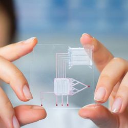
What Is an Organ-on-a-Chip?
Organ-on-a-chip platforms combine biomaterial technology, cell biology, and engineering
Clinical drug trials hinge on the success of efficacy and toxicity studies using cell culture and in vivo animals. These preclinical studies can miss drug off-target toxicity, patient subgroup sensitivities, and disease pathophysiology—highlighting the importance of developing novel approaches to health research and drug discovery. An estimated 40 percent of drugs that pass preclinical studies fail during clinical trials,1 contributing significantly to research and development costs.2 More reliable preclinical models, such as organ-on-a-chip technologies, offer a potential solution.
First coined in 2010, organ-on-a-chip (OOAC) stems from the advances made in lab-on-a-chip (LOC) technologies, while incorporating both tissue and organ-level physiology. OOAC provides a platform for disease modeling and drug testing. OOAC combines multiple cells types with engineering, such as spatial confinement of cells, sensors, and microfluidic channels. These platforms differ from organoids that rely on spontaneous self-assembly to form organ-like structures. Milica Radisic, PhD, professor at the University of Toronto and Canada Research Chair (Tier 1) in Organ-on-a-Chip Engineering, who is at the forefront of OOAC research, says “a lofty goal is to see organ-on-a-chip used in clinicals trials, keeping clinical participants safe, as well as completely eliminating the need for animal studies, which will have huge ethical and financial impact.” Recently, Radisic’s team has published an OOAC disease model used to study SARS-CoV-2 in the vasculature.3
OOAC systems aim to replicate organ mechanical stimuli and fluid flow and cell/organ crosstalk and spatial organization. Factors of organ physiology that are currently reproducible include concentration gradients, shear force, cell patterning, tissue boundaries, and tissue–organ interactions. OOAC has been widely touted as an emerging technology and has been proposed as a possible future replacement for animal testing. This article will examine the different components that constitute this fascinating technology.
Geometric confinement and cell patterning
OOAC platforms predominantly use immortalized cell lines; however, patient-derived cells, such as induced pluripotent stem cells, can improve disease modeling and enhance clinical value.4
There are two main types of OOAC tissue organization: barrier and parenchymal tissues.5 Parenchymal tissues use 3D extracellular matrices, while barrier tissues incorporate porous membranes. These membrane surfaces allow for the coculture of two cell types to recreate tissue boundaries, such as vascular and epithelial interfaces.
Biocompatible materials are crucial when building cell arrangements and act as a scaffold in shaping these arrangements and protecting cells from mechanical damage. Hydrogels and PDMS (oxygen permeable) are common biocompatible materials used in the design of OOACs. Hydrogels, while highly permeable, are also fragile.4 However, microfabrication and 3D printing can help overcome this problem. While 3D layouts closely resemble the in vivo environment, 2D arrangements are more prevalent when considering cost and time.
Microfluidics
Microfluidics technology is central to the success of OOAC platforms to transport nutrients and remove waste products from cells. In addition, the microfluidic system establishes biochemical concentration gradients and generates fluid shear, thereby inducing organ polarity. Microfluidics can also reproduce tissue and organ-level physiology such as dynamic mechanical stresses (e.g., blood and lung pressure). These periodic mechanical stresses are made using porous elastic membranes. Such mechanical stresses are essential for the differentiation of physiological processes. Active fluid flow uses micropumps that incorporate pneumatically controlled membrane deflection to circulate fluids, while a cheaper alternative is gravity-driven passive flow from reservoirs.

Environmental control
In addition to recreating the in vivo environment, the OOAC uses mechanisms to introduce chemical and physical signals necessary to simulate the physiological microenvironment.6 Such simulation includes mechanical (compressive and tensile forces), electrical (especially for neurons and muscle cells), biochemical (diffusion gradients), and spatiotemporal cues. These signals drive tissue maturation and function. An example is incorporating mechanically stretchable membranes to reproduce cyclic mechanical strains to model breathing and vascular perfusion.
Sensors
To measure the changes, OOACs incorporate built-in sensors and are often made from transparent materials to faciliate real-time microscopy. Sensors can be optical or electrochemical and are noninvasive while collecting quantitative biochemical data from the system. Examples of biosensors include physical sensing units measuring pH, oxygen, and temperature levels. In addition, more specialized sensors can measure cell metabolite concentrations and cell viability. The majority of OOACs make use of fluorescent microscopy to collect experimental data.
Moreover, incorporating biosensors into OOAC platforms removes the need for conventional analytical methods and biochemical assays that require large sample volumes and are time-consuming.7 Biosensors make use of small sample volumes, have lower detection limits, avoid disruption, are suitable for miniaturization, and allow for continuous monitoring. While biosensors are advantageous, considerable research and development are required to further integrate these sensors into OOAC platforms.
Advantages of organ-on-a-chip platforms
OOAC platforms have several advantages, such as being compatible with current imaging technologies, improving cellular fidelity, and recreating dynamic environmental factors. “The advantage of OOACs and readouts are that they are high content, enabling many sophisticated readouts to be collected from one device,” says Radisic.
Using OOAC platforms is estimated to save between 10–26 percent in research and development costs.8 These savings are driven by higher drug success rates and lower indirect costs and time during the lead optimization and preclinical phase. These cost reductions could potentially reduce market entry barriers for new biotechnology businesses.
OOAC platforms are also uniquely placed to study complex processes involved in cancer progression and treatment. For example, OOACs can replicate the acidic microenvironment of solid tumors,9 which is a current challenge in animal models. Such systems allow researchers to isolate and study the effect of pH on tumor viability.
Challenges of organ-on-a-chip platforms
While OOAC platforms are a promising technology, several challenges remain to unlock the technology’s true potential. Hurdles in biology include scaling organs, media optimization, vascularizing tissues, coculturing different tissues, managing cell density, and reproducing immune responses.
Furthermore, several technical challenges remain, such as the interaction of compounds and metabolites with bioreactors, sensor integration and biofouling, sterility, preventing bubbles, and optimizing shear and flow rates. In addition to technological challenges, regulatory hurdles also present an obstacle as they are usually based on animal models, says Radisic, adding that the novelty of this technology and the operation of OOACs are also an unfamiliar interface for biologists.
Organ-on-a-chip platforms moving forward
OOAC is a potential substitute to in vitro and animal models that often poorly reflect human physiology. Various organs have already been adapted and used in research with the ultimate goal to create a human-on-a-chip.10 Radisic’s group is already looking to introduce fractal and chaotic cues into their OOAC designs to better mimic physiological conditions. OOAC has the real potential to become an indispensable tool due to its superior nature compared to existing technologies.
References:
- Van Norman GA. Limitations of animal studies for predicting toxicity in clinical trials: Part 2: Potential alternatives to the use of animals in preclinical trials. JACC Basic Transl Sci. 2020;5(4):387-397. doi:10.1016/j.jacbts.2020.03.010
- Franzen N, van Harten WH, Retèl VP, Loskill P, van den Eijnden-van Raaij J, IJzerman M. Impact of organ-on-a-chip technology on pharmaceutical R&D costs. Drug Discov Today. 2019;24(9):1720-1724. doi:10.1016/j.drudis.2019.06.003
- Lu RXZ, Lai BFL, Rafatian N, et al. Vasculature-on-a-chip platform with innate immunity enables identification of angiopoietin-1 derived peptide as a therapeutic for SARS-CoV-2 induced inflammation. Lab Chip. 2022;22(6):1171-1186. Published 2022 Mar 15. doi:10.1039/d1lc00817j
- Zhang B, Korolj A, Lai BFL, et al. Advances in organ-on-a-chip engineering. Nat Rev Mater. 2018;3:257–278. doi.org/10.1038/s41578-018-0034-7
- Ching T, Toh YC, Hashimoto M, Zhang YS. Bridging the academia-to-industry gap: organ-on-a-chip platforms for safety and toxicology assessment. Trends Pharmacol Sci. 2021;42(9):715-728. doi:10.1016/j.tips.2021.05.007
- Ahadian S, Civitarese R, Bannerman D, et al. Organ-on-a-chip platforms: A convergence of advanced materials, cells, and microscale technologies [published correction appears in Adv Healthc Mater. 2018 Jul;7(14):]. Adv Healthc Mater. 2018;7(2):10.1002/adhm.201700506. doi:10.1002/adhm.201700506
- Rodrigues RO, Sousa PC, Gaspar J, Bañobre-López M, Lima R, Minas G. Organ-on-a-chip: A preclinical microfluidic platform for the progress of nanomedicine. Small. 2020;16(51):e2003517. doi:10.1002/smll.202003517
- Ma C, Peng Y, Li H, Chen W. Organ-on-a-chip: A new paradigm for drug development. Trends Pharmacol Sci. 2021;42(2):119-133. doi:10.1016/j.tips.2020.11.009
- Lam SF, Bishop KW, Mintz R, Fang L, Achilefu S. Calcium carbonate nanoparticles stimulate cancer cell reprogramming to suppress tumor growth and invasion in an organ-on-a-chip system. Sci Rep. 2021;11(1):9246. doi:10.1038/s41598-021-88687-6
- Wu Q, Liu J, Wang X, et al. Organ-on-a-chip: recent breakthroughs and future prospects. Biomed Eng Online. 2020;19(1):9. doi:10.1186/s12938-020-0752-0


