
A Timeline of the CRISPR/Cas System
From discovery and development to clinical applications

Michelle Dotzert is the creative services manager for our partner brand, Lab Manager. She holds a PhD in kinesiology (specializing in exercise biochemistry) from the University of Western Ontario. Her research examined the effects of exercise training on skeletal muscle lipid metabolism and insulin resistance in a rodent model of Type 1 Diabetes. She has experience with a variety of molecular and biochemistry techniques, as well as HPLC-MS.
ViewFull ProfileLearn about ourEditorial Policies.
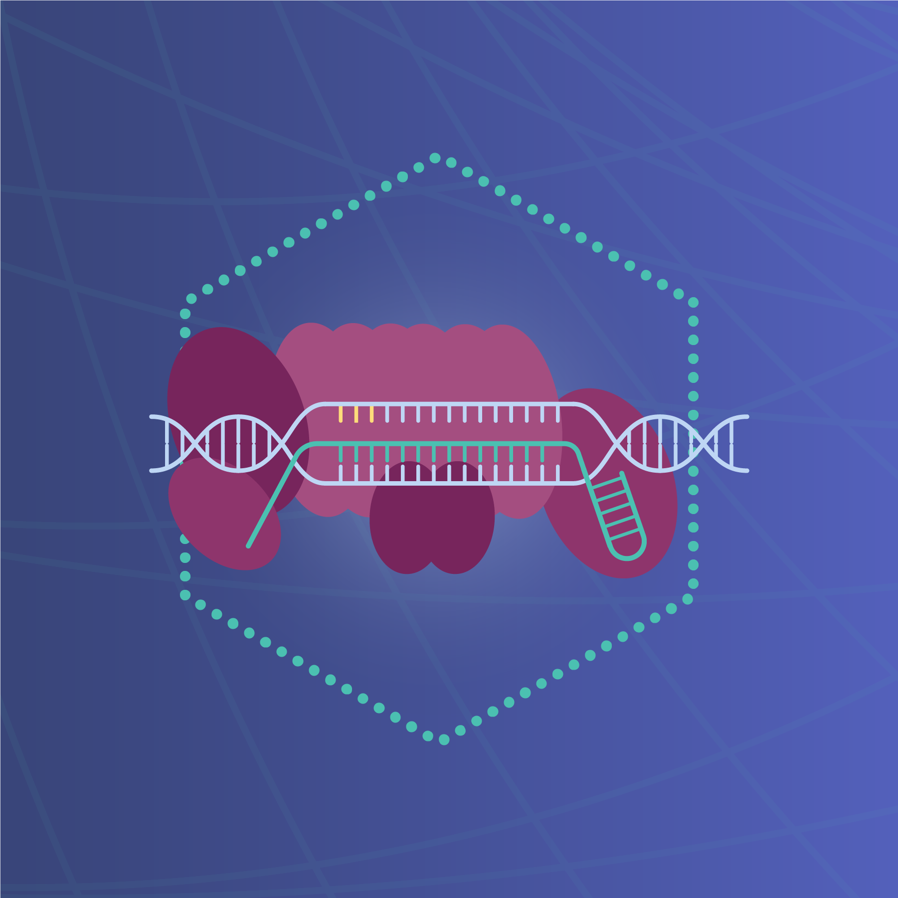
The Clustered Regularly Interspaced Short Palindromic Repeats/CRISPR-associated protein systems are bacterial and archaeal adaptive immunity systems, capable of protecting cells from invading nucleic acids. Of these, the CRISPR/Cas9 system is used for genome editing, as the Cas9 protein targets and binds specific DNA sequences. There are three types of CRISPR systems, each consisting of CRISPR-associated genes, noncoding RNAs, and an array of repetitive elements interspaced with protospacers (CRISPR RNA [crRNA]). Each protospacer within the DNA target is associated with a protospacer adjacent motif (PAM). The Type II CRISPR system, CRISPR/Cas9, consists of the Cas9 nuclease, a crRNA array encoding guide RNAs, and an auxiliary trans-activating crRNA (tracrRNA). Fusing the crRNA and tracrRNA creates a single-guide RNA (sgRNA), capable of directing Cas9 toward a target within the vicinity of the PAM sequence.
When the guide sequence pairs with the DNA target, Cas9 mediates a double-stranded break upstream of the PAM. This initiates DNA repair via non-homologous end joining (NHEJ) or homology-directed (HDR) repair pathways. Without a repair template, the NHEJ process creates insertion/deletion mutations, and can be used to knock out specific genes. Alternatively, with the introduction of an exogenous repair template, the HDR pathway can be used to create modifications at a specific target locus.

1987
In the process of analyzing the E. coli genome for genes associated with phosphate metabolism, researchers made an unexpected discovery. They found five homologous sequences of 29 nucleotides arranged in direct repeats with 32 nucleotides as spacing in the 3’- end flanking region of the iap gene. This repetitive sequence was later defined as CRISPR.

1993
Halophilic archaea, such as Haloferax mediterranei, are able to adapt to environmental changes in salinity. While studying genetically uncharacterized regions of the H. mediterranei genome, scientists discovered 30-bp long DNA segments, repeated at regular distances. Further sequencing experiments confirmed the region contained at least 14 near-perfectly conserved repeats flanked at one end by a highly degenerated copy.

2002
Using in silico analysis, four CRISPR-associated (Cas) genes were identified in CRISPR-containing prokaryotes, each located adjacent to a CRISPR locus. These genes were absent in CRISPR-negative prokaryotes.

2005
Three independent research groups demonstrated sequence similarity between spacer regions of CRISPRs and sequences of bacteriophages and plasmids. Species containing CRISPR elements were protected from corresponding foreign invaders, demonstrating its function as an immune system.
At the same time, a motif necessary for CRISPR to function as an immune system was first discovered in Streptococcus strains. Later, other motifs were defined for organisms with different CRISPR systems. These short sequence motifs adjoining protospacers were later termed PAM (protospacer adjacent motif).

2007
The CRISPR/Cas system is demonstrated to function as an immune system in an experiment with a phage-host model system. A phagesensitive wild-type S. thermophilus strain and two virulent bacteriophages were selected, and several phageresistant mutants were generated by challenging the wild-type strain with the phages. Novel spacers derived from phage DNA were integrated into the CRISPR1 locus, conveying resistance to the corresponding phage. When the protospacer sequences were removed, resistance disappeared.
2008
The molecular mechanisms underlying the CRISPR-based defense system had yet to be determined. Using Cas protein tagging and affinity purification, a protein complex of five Cas proteins (CasA, CasB, CasC, CasD, and CasE) was identified. The complex, Cascade (CRISPR-associated complex for antiviral defense) was isolated from E. coli lysates. The CRISPR RNA endonuclease subunit of Cascade forms mature guide RNAs that are essential for antiviral defense.

2012
In vitro studies demonstrated that the Cas9-crRNA complex functions as an RNA-guided endonuclease, with Cas9 causing DNA cleavage, and that crRNA base-paired to tracrRNA also directs Cas9 to introduce double-stranded breaks.

2013
The ability to edit the genome of eukaryotic cells using CRISPR/Cas9 was demonstrated with targeted genome cleavage in human and mouse cells.
CRISPR-on (nuclease-dead Cas9 protein fused with a transcriptional activation domain and sgRNA) is shown to effectively activate reporter genes in human and mouse cells.
Scientists demonstrated an imaging technique using enhanced green fluorescent protein (EGFP)-tagged endonuclease-deficient Cas9 protein and sgRNA to image repetitive elements in telomeres and coding genes in living cells.
Scientists were able to rescue mice with a dominant mutation in the Crygc gene that causes cataracts, by injecting Cas9 mRNA and sgRNA into zygotes.

2014
Cas9/sgRNA screens were established as a tool for systematic genetic analysis in mammalian cells. The CRISPR/Cas9 system has several potential benefits over existing functional screening methods, including the ability to study phenotypes that require a complete loss of function gene to be elicited, a low rate of false negatives in largescale screens, and the lack of effect of off-target effects on screens.
The genome engineering capabilities of CRISPR/Cas9 were demonstrated in primates, with successful targeting of several genes in monkey embryos by co-injection of Cas9 mRNA and sgRNAs. The CRISPR/Cas9 system enabled disruption of Ppar-γ and Rag1 genes with no off-target mutagenesis.

2015
The first study demonstrating gene editing of human embryos was published. Scientists used CRISPR/Cas9 to cleave the endogenous β-globin gene in tripronuclear zygotes. Despite effectively cleaving the gene, HDR was low, off-target cleavage was observed, and the endogenous delta-globin gene (homologous to the β-globin gene) competed with exogenous oligos as the repair template which led to mutations.
Scientists developed a new approach to modeling human liver disease in mice by performing in vivo CRISPR/Cas9 genome editing. They administered a Streptococcus pyogenes-derived Cas9 system (SpCas9) targeting Pten, a gene involved in non-alcoholic steatohepatitis (NASH). Months later, the mice demonstrated characteristics of NASH consistent with models generated using the Cre-loxP system.

2016
Scientists used CRISPR/Cas9 to improve chimeric antigen receptor (CAR) T-cell therapies by reducing alloreactivity. Disrupting multiple genomic loci led to the production of CAR T cells deficient in the expression of endogenous T-cell receptor (TCR) and HLA (human leukocyte antigen) class I, thereby reducing alloreactivity and preventing graft-versus-host disease.
Targeted DNA methylation editing was achieved. Fusion of Tet1 (teneleven translocation methylcytosine dioxygenase 1) or Dnmt3a (DNA (cytosine-5)-methyltransferase 3a) enzymes with a catalytically inactive Cas9 enables targeted DNA methylation editing. Targeting the fusion protein caused activation or silencing of an endogenous reporter.

2017
RNA base editing technology was developed using type VI CRISPR-associated RNase Cas13. Scientists engineered the REPAIR system (RNA Editing for Programmable A to I Replacement), to edit full-length transcripts containing pathogenic mutations. A catalytically inactive Cas13 directed adenosine-to-inosine deaminase activity by ADAR2 (adenosine deaminase acting on RNA type 2) to transcripts in mammalian cells.
2018
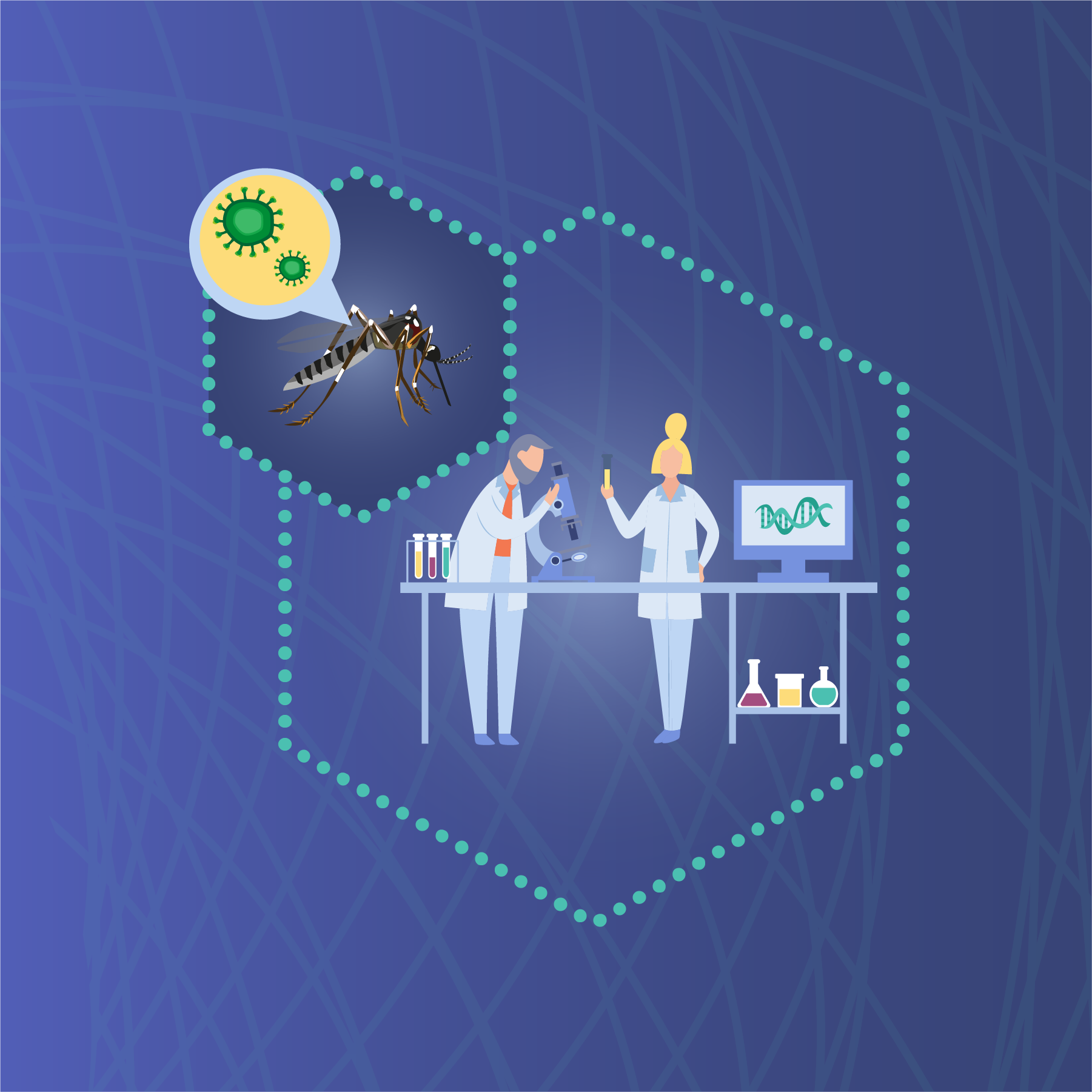
Scientists developed a viral diagnostic platform using Cas13. SHERLOCK (Specific High-sensitivity Enzymatic Reporter un-LOCKing) amplifies RNA, and combines amplified nucleotides with Cas13a, a guide RNA, and a nucleotide sequence coupled to a fluorescent reporter and quencher. If the target sequence is present, Cas13a will cleave the RNA reporter and activate the fluorophore. SHERLOCK has been shown to detect Zika virus and dengue virus in patient samples.
Researchers in the United Kingdom published a paper demonstrating potential risks of CRISPR/Cas9 technology. Long-read sequencing and long-range PCR genotyping of mouse embryonic stem cells, hematopoietic progenitor cells, and a human differentiated cell line revealed sgRNA/Cas9 induced DNA breaks often resolved into deletions over many kilobases. Results suggested potential pathogenic consequences of gene editing.

2019
A new gene editing system was characterized and engineered. ShCAST (CRISPR-associated transposase from cyanobacteria Scytonema hofmanni) consists of Tn7-like transposase subunits and the type V-K CRISPR effector (Cas12k). It catalyzes RNA-guided DNA transposition, and was used to integrate DNA into targeted sites in the E. coli genome.

2020
In early 2020, an outbreak of a novel respiratory virus, SARS-CoV-2 (severe acute respiratory syndrome coronavirus 2) rapidly evolved into a global pandemic. One of the greatest challenges was to develop a rapid, accurate testing method that could be used to identify infectious individuals and reduce disease transmission. A CRISPR-Cas13a-based assay was developed for the detection and quantification of SARS-CoV-2 RNA. Unlike other assays, this approach enables direct detection of RNA without any additional manipulations such as amplification. A single guide RNA is used to detect viral RNA, and a fluorescent signal may be measured with a mobile phone camera. As such, it is a potentially fast, accurate, and portable option for SARS-CoV-2 screening.